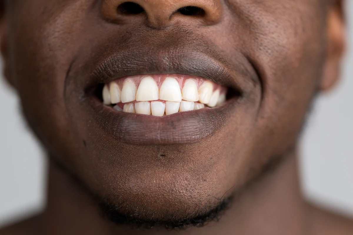
Dentistry has lived for decades with an awkward asymmetry: bone heals, skin closes, and the hardest tissue in the human body does neither. Once a tooth finishes developing, the cells that built its enamel are gone, so lost enamel stays lost. Work from the University of Nottingham, reported in Nature Communications on 5 November, does not reverse that biology. It routes around it, using a material to perform the scaffolding that cells once did, and in doing so it challenges the practical conclusion dentists have had to deliver to patients for years.
Saliva already contains calcium and phosphate, and it can put back a small amount of mineral lost in the very earliest stages of demineralisation. That is the honest basis for remineralising products. But the quantities involved are trivial compared with actual tissue loss: once acid erosion or abrasion has removed a meaningful layer, saliva can slow further damage but cannot rebuild the missing architecture. Fluoride varnishes and similar treatments make surviving enamel more acid-resistant and can arrest early chalky lesions. They are protective measures, and every dentist knows the point at which protection stops and a filling, veneer or crown begins.
Saliva already contains calcium and phosphate, and it can put back a small amount of mineral lost in the very earliest s
The Nottingham group, drawn from the School of Pharmacy and the Department of Chemical and Environmental Engineering, inverted the usual question. Rather than delivering more mineral to the tooth, they asked how mineral ought to be arranged. Their material is a protein-based gel designed to imitate key features of the natural matrix proteins that guide enamel formation during tooth development. It contains no fluoride. Its job is not to supply building blocks but to supply order, which turns out to be the harder and more consequential half of the problem.
Application was deliberately kept close to existing practice: a clinician can paint the gel onto teeth much as a fluoride varnish is applied, with no new equipment, no curing step and no elaborate preparation. This looks like a modest design choice and is actually decisive for adoption. Any extra minutes added to an appointment reduce the likelihood that a technique reaches routine care, and a laboratory material that demands a completely new workflow tends to stay in the laboratory regardless of how well it performs.
Once applied, the gel forms a thin durable coating that penetrates the surface and fills microscopic cracks and gaps. That solves attachment first — a film merely resting on enamel would be stripped by the first few chewing cycles — and then provides a framework for mineral growth anchored to the existing tooth. The coating attracts calcium and phosphate ions naturally present in saliva and organises them into new crystals. The researchers describe this as epitaxial mineralisation: new crystals grow in alignment with the old ones, so the regenerated material becomes structurally continuous with the original surface rather than sitting on top of it as a separate layer.
Order matters more than deposition because enamel derives its hardness and durability from the precise arrangement of its crystals, not simply from what it is made of. Scattering mineral particles onto a tooth would produce a brittle coating with nothing like the mechanical behaviour of the real tissue. Lead author Abshar Hasan, a postdoctoral fellow, frames the achievement in exactly these terms: the material promotes integrated, organised crystal growth that recovers the architecture of healthy natural enamel. Architecture before composition is the line that separates this work from earlier remineralisation attempts.
The team then tested whether that architecture survives use, measuring mechanical behaviour under conditions simulating brushing, chewing and exposure to acidic foods. The regenerated tissue performed comparably to healthy natural enamel, which suggests a load-bearing layer rather than a temporary protective film. A related result extends the approach to exposed dentine, the softer tissue beneath enamel whose microscopic channels transmit hot, cold, sweet and acidic sensations toward the nerve. Growing an enamel-like mineral layer there could reduce sensitivity and improve how fillings and other restorations bond to the tooth.
Translation is already underway. Professor Alvaro Mata, who led the study, stresses that the technology was designed with the clinician and patient in mind: safe, quick to apply, scalable, and versatile enough to appear in several product formats. The group has begun commercialisation through the start-up Mintech-Bio with the aim of a first product the following year. Enamel deterioration contributes to dental problems affecting close to half of the world's population, and oral health is linked to wider conditions including diabetes and cardiovascular disease, so moving intervention earlier in the sequence would matter. Still, the evidence here is laboratory mechanical testing rather than long-term patient follow-up: durability under continuous acid challenge, variation in saliva composition between individuals, and performance across a real dentition all remain open questions. The direction has changed; routine care has not caught up yet.


Comments