
The search for ageing biomarkers has a familiar rhythm: pick a molecule, watch it rise or fall with age, then argue about whether it causes anything or merely reports damage. DNA lesions, telomeres, inflammation, proteostasis — all have been worked over. A new study in Nature Aging asks a different question: what if the signal is not one molecule going up, but an entire distribution quietly sliding sideways?
The distribution in question belongs to lipid acyl chains. A glycerophospholipid is roughly a polar head plus two hydrophobic tails, and the number of carbons and double bonds in those tails sets membrane thickness, fluidity and how comfortably embedded proteins do their jobs. Two enzyme systems govern the length: ELOVL family elongases extend chains, beta-oxidation shortens them. Until now, nobody had mapped how chain length shifts with age across tissues and species in a systematic way.
The group used UPLC-HRMS lipidomics and, crucially, turned a visual impression into a statistic. For each lipid class they computed the Pearson correlation between total acyl-chain carbon count and the fold change with age, then used a one-sample Wilcoxon test to ask whether a tissue shows a global shift. The result is a reusable readout: not 'this looks longer', but a number that can be compared across datasets and, later, across interventions.
The counterintuitive half of the finding is depletion, not accumulation. In 9 of 16 ageing datasets, more than half of the lipid classes that lengthened also lost short- and medium-chain species; the inflection clusters around C18. Ageing lipid remodelling looks like a redistribution rather than a simple pile-up of long molecules. Double-bond counts rise alongside chain length, so overall unsaturation balance is largely preserved — the pattern reads as directed rewriting, not random decay.

Conservation is documented densely. Six of ten mouse tissues show statistically significant global lengthening, including heart, liver, kidney, spleen, gastrocnemius and brown fat. Ageing C. elegans and Drosophila show the same trend. In humans, skeletal muscle cohorts comparing 20–30 with 65–80 year olds, and heart biopsy cohorts spanning younger and older men and women, both show age-associated lengthening, consistent across sexes.
One easily missed clue points beyond the clock itself: in a heart disease progression cohort, the positive association between chain length and age strengthens as hypertrophic cardiomyopathy worsens from grade 1 to grade 2. That hints the readout tracks pathological organ damage as well as chronological ageing, though the paper carefully leaves it at correlation and does not offer a diagnostic test.
So can the classic anti-ageing interventions reverse it? The team ran comparisons in cultured cells, living animals and a human cohort, and the answers came apart. Rapamycin shortened chains globally in HL-1 cardiac cells but, given intermittently to living mice, moved only a few lipid subclasses. Four days of one-hour daily high-intensity exercise in postmenopausal women adjusted chain length only in cholesteryl esters and triglycerides.
Dietary restriction — 60% of ad libitum intake — was the only intervention under the tested conditions that reliably shortened chains across the whole lipidome, in the hearts of both young and old female mice. Headlines reduced this to 'eating less really does slow ageing', which is directionally fair but flattens the most useful part of the work. The real lesson is the in-vitro versus in-vivo gap: a candidate that looks convincing in a cell line may do something far narrower in an organism.
By the same logic, 'exercise lost' must not be read as 'exercise does not help'. The human arm lasted four days and measured one narrow omics endpoint; the paper explicitly states that long-term exercise was not assessed and that the result does not diminish exercise's established health value. A negative result on one endpoint and a verdict on a lifestyle are different claims at different levels, and stating the boundary that clearly makes the study more trustworthy, not less.
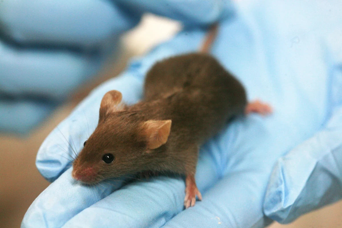
Mechanistically, the work lands on Plb1, which encodes phospholipase B. In BXD genetically diverse mouse strains, muscle Plb1 expression correlates negatively with lifespan. Human Mendelian randomisation links genetically predicted higher PLB1 expression to a higher frailty index, with no significant association to heart failure. In worms, seven of nine orthologues were amenable to RNAi and all seven extended lifespan; knocking down F36A2.9 shortened chains globally, and supplementing C18:1, C20:1 or C22:1 long-chain fatty acids abolished the longevity gain, which depends on daf-16 and skn-1. Here the causal chain actually closes.
That target moves the field's gaze. Most ageing lipid work has watched the synthesis end, especially ELOVL elongases. Plb1 belongs to the disassembly and recycling side — beta-oxidation, Lands cycle, phospholipases. A distribution is set by both production and removal, so watching only one end leaves half the equation unwritten.
The study also dismantles the tempting slogan that shorter is always better. Knocking down elo-3, another elongase, did not globally shorten cardiolipin or BMP species the way elo-1 knockdown did, and it shortened lifespan instead. Molecular dynamics simulations revise the mechanism too: in single-component artificial membranes, longer chains do thicken the bilayer, but real membranes are complex mixtures, and even 25% chain elongation changes bulk physical parameters only modestly. The authors suspect local microenvironments, disrupted membrane-associated proteins and greater susceptibility to lipid peroxidation, rather than a membrane that suddenly goes rigid.
The limits deserve the last word. Quantification is relative between groups, not absolute abundance; causal evidence rests largely on worms; human tissue supports correlation only, with no intervention trial; and Mendelian randomisation used whole-blood eQTL as a proxy for gene expression in the target organ. None of this is a dietary recommendation — and the chain length of fat in food is not the chain length of lipids in tissue. What the work leaves behind is better than advice: a quantitative omics endpoint, so that screening for dietary-restriction mimetics finally has something measurable to aim at.





Comments